
Your skeleton is not a single, solid lump of bone. It is a complex framework of over 200 individual bones, and for that framework to function, the bones must connect. The points where they meet are called joints.
Some joints, like those in your shoulder or knee, allow sweeping, dynamic movement. Others are locked tight, stitching your skull together so firmly that they appear to be one piece. Somewhere in the middle lies a fascinating category: joints that allow a little give but never move freely. These are the cartilaginous joints.
If you are studying for a GCSE, A-level, or introductory anatomy exam in the UK, these joints will appear in your curriculum. They are also clinically important in understanding back pain, growth development, and pelvic stability. Let’s break down exactly what they are, how they work, and why they matter.
What Are Cartilaginous Joints?
Cartilaginous joints are connections between bones held together entirely by cartilage. They allow only limited movement and contain no joint cavity.
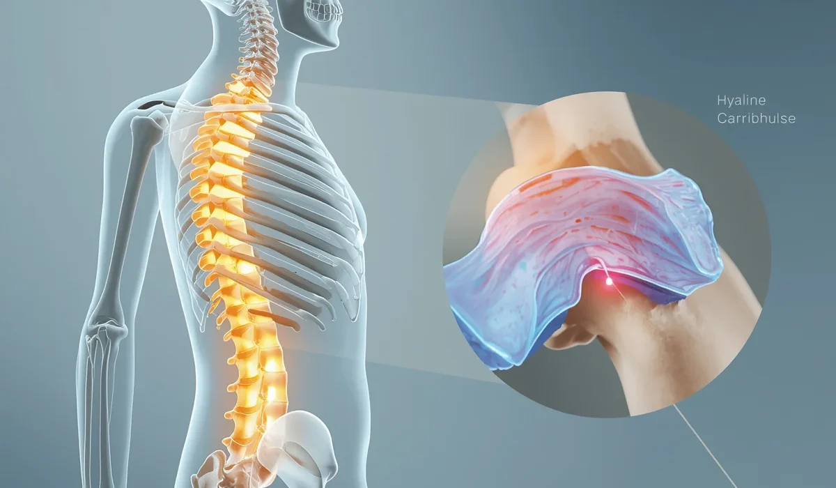
Unlike a knee joint, which has a fluid-filled capsule and slippery surfaces designed for free motion, a cartilaginous joint is simpler. The bones are linked by a bridge or disc of cartilage—either tough, glassy hyaline cartilage or densely packed fibrocartilage. This tissue acts as a cushion and a connector simultaneously.
Because there is no lubricating synovial fluid and no spacious cavity, these joints are sometimes called amphiarthroses, which translates roughly to “both sides joint”—meaning they sit halfway between rigid and freely movable .
These joints are vital for two contradictory roles: they must hold bones together firmly while still allowing a small degree of give. Without them, your spine couldn’t absorb the shock of walking, and your pelvis couldn’t widen safely during childbirth.
What Are the Types of Cartilaginous Joints?
The UK anatomy curriculum splits cartilaginous joints into two distinct types based on the kind of cartilage present and whether the joint is designed to be temporary or permanent.
Primary Cartilaginous Joints (Synchondrosis)
A primary cartilaginous joint, or synchondrosis (pronounced sin-con-dro-sis), is a joint where bones are connected exclusively by hyaline cartilage.
Hyaline cartilage is smooth, glassy, and firm. In a synchondrosis, it acts like a rigid glue. This type of joint permits almost no movement at all; it is designed for stability and, often, for eventual bone growth.
The defining characteristic of most primary cartilaginous joints is that they are temporary. They exist to allow a skeleton to grow longer, and once growth stops, the cartilage gradually ossifies and turns into solid bone . When this happens, the joint effectively disappears.
There is one notable exception. The joint between the first rib and the sternum (the costochondral joint) is a permanent synchondrosis. The hyaline cartilage there remains intact throughout adulthood because that rib needs to stay securely anchored while the rest of the ribcage moves during breathing .
Secondary Cartilaginous Joints (Symphysis)
A secondary cartilaginous joint, or symphysis (pronounced sim-fih-sis), is a joint where bones are covered by a thin layer of hyaline cartilage but are predominantly connected by a thick pad of fibrocartilage.
Fibrocartilage is tougher and more fibrous than the hyaline type, packed with strong collagen bundles. This makes a symphysis exceptionally good at absorbing compressive forces.
Crucially, these joints are permanent. They are built to last a lifetime, resisting immense daily pressure without wearing out under healthy conditions. They allow a limited but functionally important degree of movement, often described as give or flexibility rather than obvious motion .
What Is the Difference Between Synchondrosis and Symphysis?
This is a classic GCSE and A-level examination question. The simplest comparison looks like this:
– Synchondrosis (Primary): Made only of hyaline cartilage. Usually ossifies and disappears once growth is complete. Allows virtually no movement.
– Symphysis (Secondary): Has both hyaline cartilage layers and a central fibrocartilage disc. Is permanent. Allows slight, cushioned movement.
If you visualise a synchondrosis, think of a temporary scaffold that hardens into the main structure. Visualise a symphysis and think of a permanent shock absorber, like a tough rubber cushion that never turns to stone .
From a functional perspective, the synchondrosis is strictly about structural growth; the symphysis is about lifelong mechanical support.
Where Are Cartilaginous Joints Found in the Body?
Cartilaginous joints are found almost exclusively along the midline of the body, where central stability is most needed.
The two most significant and commonly cited locations in the human skeleton joints UK curriculum are the spinal column and the pelvis. You will also find synchondroses in the long bones of a growing child’s limbs .
What Are Examples of Cartilaginous Joints?
Here are the key examples you need to remember, categorised by joint type . Each illustrates a different core function of cartilaginous joints.
Synchondrosis Examples
– Epiphyseal (Growth) Plates: Found near the ends of long bones like the femur and humerus in children and adolescents. These hyaline cartilage plates allow the bone shaft to lengthen. Once you reach your final adult height, hormonal changes trigger ossification, and the plate becomes a permanent bony line .
– First Sternocostal Joint: The connection between the first rib and the sternum. This synchondrosis uniquely does not ossify, providing a rigid anchor that keeps the upper ribcage stable during breathing .
– Spheno-occipital Synchondrosis: A less commonly cited example found at the base of the skull. This cartilage joint between the sphenoid and occipital bones fuses fully during adolescence .
Symphysis Examples
– Intervertebral Discs: The pads of fibrocartilage wedged between the vertebrae of your spine. Each disc bonds firmly to the vertebral bodies above and below. Collectively, the 23 discs make up about a quarter of your spinal column’s total length and are the most functionally important cartilaginous joints in the body .
– Pubic Symphysis: The joint at the front of the pelvis where the two pubic bones meet. A thick fibrocartilage disc sits between them, reinforced by strong ligaments. This joint normally permits very little movement in men, but in women during pregnancy, hormonal changes soften the cartilage to allow slight widening for childbirth .
– Manubriosternal Joint: The joint between the upper portion of the sternum (manubrium) and the main body of the sternum. This symphysis often partially ossifies with age but retains enough fibrocartilage to allow slight movement during breathing.
– Sacroiliac Joints (mixed classification): Some textbooks classify the sacroiliac joints as partially cartilaginous (symphysis) and partially fibrous, but they are increasingly taught under symphysis characteristics in UK syllabuses .
What Is the Function of Cartilaginous Joints?
The function of cartilaginous joints can be summed up in three essential roles: they provide structural stability, they absorb mechanical shock, and they permit a very limited, controlled degree of movement.
The spine is the clearest example. When you walk, run, or jump, the intervertebral discs compress slightly, flattening out to absorb impact before springing back. Without this cartilaginous mechanism, the vertebrae would grind against each other, causing rapid bone damage and nerve impingement.
At the pubic symphysis, the function is different but equally important. It locks the left and right sides of the pelvis together to withstand the forces of standing and walking, yet it allows just enough compliance to flex under load rather than fracture .
In a growing skeleton, the function of a synchondrosis is radically different: it provides a structurally sound framework that actively divides, enabling longitudinal bone growth until skeletal maturity .
Are Cartilaginous Joints Movable?
Cartilaginous joints are slightly movable (amphiarthrotic). They allow only a small amount of give or compression, not free movement.
This is a common confusion point. You cannot voluntarily “move” a symphysis the way you flex an elbow. The movement at a cartilaginous joint is passive and measured in degrees of compression and slight tilting rather than visible range of motion.
For instance, your intervertebral discs allow you to bend your spine forward, but that bending is a sum of very small movements occurring across many adjacent joints simultaneously. No single disc contributes more than a sliver of tilt. Likewise, the pubic symphysis can rotate slightly during walking, but you would never describe it as a mobile joint.
This slight mobility is what makes these joints uniquely vulnerable to degenerative wear. They sit in a biomechanical middle ground: rigid enough to bear weight, pliable enough to flex, but susceptible to gradual breakdown over decades .
Comparison: Fibrous vs Cartilaginous vs Synovial Joints
For UK students tackling anatomy exams, a structured comparison table is invaluable. Here is the simplified version of what you need to know .
| Feature | Fibrous Joints | Cartilaginous Joints | Synovial Joints |
|---|---|---|---|
| Connecting Material | Dense fibrous connective tissue (collagen) | Hyaline cartilage or fibrocartilage | Articular cartilage with synovial fluid in a capsule |
| Joint Cavity | Absent | Absent | Present |
| Movement Level | Immovable (synarthrosis) | Slightly movable (amphiarthrosis) | Freely movable (diarthrosis) |
| Key Examples | Skull sutures, teeth sockets | Intervertebral discs, pubic symphysis, growth plates | Knee, hip, shoulder, elbow |
| Permanence | Permanent (mostly) | Primary often temporary; secondary permanent | Permanent |
Fibrous joints lock things down. Synovial joints free things up. Cartilaginous joints occupy the middle territory, providing exactly the right balance of stability and cushioning.
Real-Life Importance for Movement and Health
The relevance of cartilaginous joints extends well beyond textbooks. They are central to everyday comfort, athletic performance, and long-term mobility.
Consider your morning routine. When you get out of bed and stretch, your intervertebral discs have rehydrated slightly overnight, making your spine feel stiff until the fluid redistributes. When you walk downstairs, the discs compress and cushion the load. When a pregnant woman’s body releases the hormone relaxin, it softens the fibrocartilage of the pubic symphysis, allowing the pelvis to adapt for birth—a remarkable demonstration of a previously rigid joint becoming temporarily more pliable .
Even in fitness, understanding these joints matters. Weightlifters rely on proper spinal alignment to protect their discs from herniation under heavy loads. A herniated disc occurs when the tough outer layer of fibrocartilage tears, allowing the softer inner material to bulge out and press against spinal nerves . This is a direct injury to a secondary cartilaginous joint.
Common Issues Affecting Cartilaginous Joints
Because cartilaginous joints have no direct blood supply to their inner cartilage, they heal very slowly when damaged.
Intervertebral disc degeneration is one of the most common musculoskeletal complaints in the UK. With age, the fibrocartilage discs lose water content, becoming thinner and less elastic. This reduces the space between vertebrae and can lead to chronic lower back pain.
In younger athletes or those with poor lifting technique, a disc can herniate acutely, causing sciatica when it compresses the nearby nerve roots.
At the pubic symphysis, dysfunction can occur during pregnancy or after trauma, causing significant pelvic pain. In some cases, the joint widens too much (diastasis), requiring physiotherapy to stabilise the pelvis.
For primary cartilaginous joints, a fracture through a child’s growth plate (epiphyseal fracture) is a serious injury. Because the hyaline cartilage is structurally weaker than the surrounding bone, a fracture can disrupt future bone growth if not correctly treated .
Why Cartilaginous Joints Matter
Cartilaginous joints are the quiet workhorses of the human skeleton. They do not generate the headline-grabbing range of motion that a shoulder does. Instead, they perform the less glamorous but more fundamental task of holding the body’s central axis together while absorbing the relentless compressive forces of daily life.
For UK students, mastering this topic means being able to clearly distinguish between primary and secondary types, giving precise anatomical examples, and explaining the functional trade-off between stability and slight mobility. For everyone else, it means understanding why your spine feels stiff in the morning, why your pelvis needs to be both rock-solid and slightly flexible, and why good posture genuinely protects your long-term health.
Whether you are revising for an exam or simply curious about how your body works, the humble cartilaginous joint deserves considerably more credit than it usually receives. Follow UKHealthInsight for more tips!